
✨ Nursing Note A&P – Tissue Level of Organization✨
Tissue Level of Organization

Four tissue types
Epithelial tissue: exposed surfaces, lines internal passageways and chambers, forms glands.
Connective tissue: fills internal spaces, provides structural support for other tissues, transports materials within the body, stores energy.
Muscle tissue: specialized for contraction and includes the skeletal muscles of the body, muscle of the heart, muscular walls of hollow organs.
Neural tissue: carries information from one part of the body to another in the form of electrical impulses.
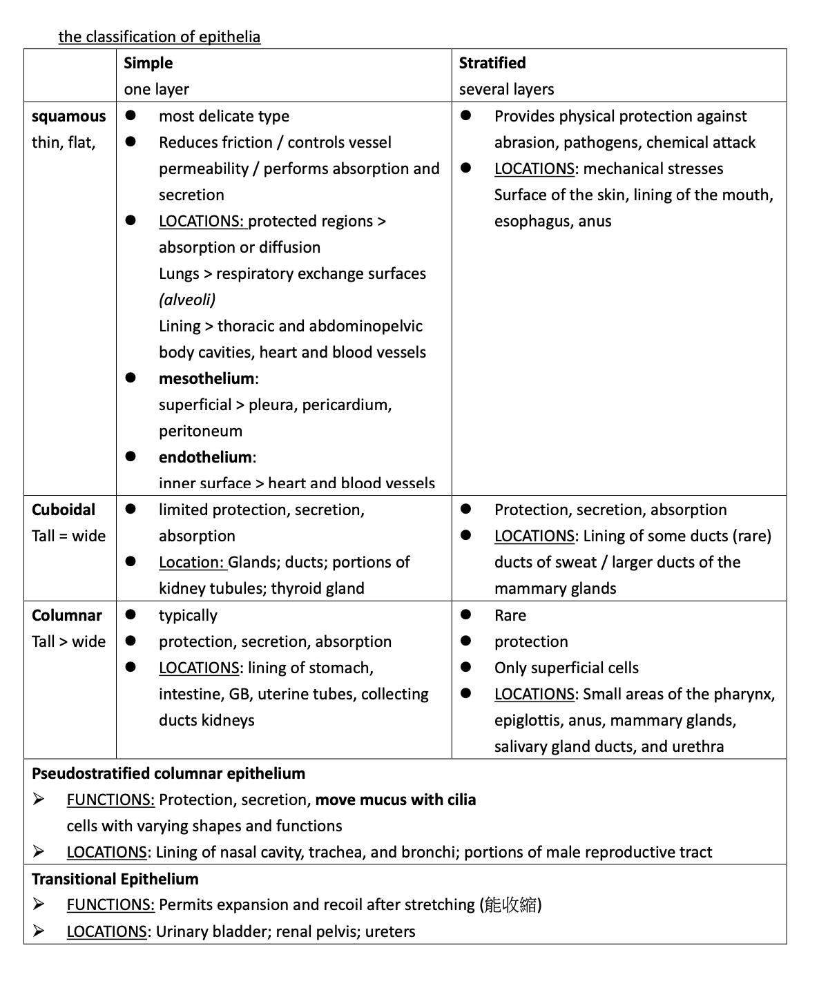
Epithelial tissue
Epithelia > cover internal or external surfaces (skin and line the digestive, respiratory, reproductive, urinary tracts)
Glands > produce fluid secretions
Function of epithelial tissue
Provide physical protection > maintain BT, blood fluid, against outside environment, infection, harm
Control permeability
Provide sensation > light tough, pressure, pain, thermoregulation
Produce specialized secretions > glandular epithelium (gland cells)
Characteristics of epithelia
Polarity > apical and basal surfaces attached
Cellularity > cell junctions
Attachment: basal lamina (base epithelium) > bound to a thin, noncellular basement membrane > complex structure (basal surface epithelium and the underlying connective tissue)
Avascularity > lack blood vessels, get nutrients by diffusion, absorption
Regeneration > repair via stem cells
Specializations of epithelial Cells
providing protection and lubrication > movement of fluids over the epithelial surface
control permeability > movement of fluids through the epithelium
production of secretions > provide physical protection or act as chemical messengers
Microvilli: absorption and secretion > digestive system, kidneys
Cilia: synchronized beating of the cilia moves substances over the epithelial surface. > respiratory tract
Maintaining the integrity of epithelia
Cell adhesion molecules (CAMs) > bind to each other and to extracellular materials.
Gap junctions
two cells are held together by two interlocking transmembrane proteins
> connexons: contain > six connexin proteins, cylinder with a central pore (narrow passageway lets small molecules and ions pass between the cells)
LOCATIONS: cardiac muscle tissue and smooth muscle tissue
Tight junction (occluding junction)
CONTAIN: two lipid plasma membranes are tightly bound together by interlocking membrane proteins
FUNCTION: prevent water and solutes from passing between the cells
stretching, bending, twisting, compression > protect mechanical stresses
Desmosome
CONTAIN: CAMs and proteoglycans bind together
FUNCTION: very strong and can resist stretching and twisting
Glandular epithelia
produce secretions
Endocrine glands (ductless glands)
FUNCTION: produces endocrine secretions (hormones) > release into interstitial > enter the bloodstream >> body.
ductless: not released into ducts
E.g., pancreas, thyroid gland, thymus, pituitary gland
Exocrine glands
FUNCTION: produce exocrine secretions > release onto an epithelial surface through tubular ducts
E.g., digestive tract, perspiration (skin), tears (eyes), milk (mammary glands)
Serous glands > parotid salivary glands
Mucous glands > sublingual salivary glands, submucosal glands(small intestine)
Mixed exocrine glands contain > submandibular salivary glands
Modes of Secretion
Merocrine secretion (common): released from secretory vesicles by exocytosis
Mucin: mixes with water to form mucus > lubricant, a protective barrier, a sticky trap for foreign particles and microorganisms
mucous (salivary glands): coat food and reduce friction during swallowing
sweat (merocrine sweat glands): produce the watery perspiration that helps cool on a hot day
Apocrine secretion: loss of cytoplasm when secretory product
apical portion of the cytoplasm> becomes packed with secretory vesicles and is then shed.
Milk production (mammary glands): merocrine and apocrine secretions
Holocrine secretion: destroys the whole gland cell when secretory product
Sebaceous glands (hair follicles)
Connective Tissues
Classification of Connective tissues
Connective tissue proper:
connective tissues with many types of cells and extracellular fibers in a syrupy ground substance.
loose connective tissues / adipose tissue / fat
dense connective tissues / tendons
Fluid connective tissues:
blood
lymph
watery matrix that contains dissolved proteins
Supporting connective tissues:
cartilage (gel with characteristics)
bone (mineral deposits)
less diverse cell population
matrix containing much more densely packed fibers
protect soft tissues and support the weight of part or body
Function of connective tissue
Establishing a structural framework for the body.
Transporting fluids and dissolved materials.
Protecting delicate organs.
Supporting, surrounding, and interconnecting other types of tissue.
Storing energy (triglycerides)
Defending body from invading microorganisms.
Cells of connective tissue proper
Fibroblasts: most abundant
secrete > hyaluronan (lock epithelial cells together) and proteins.
also secretes protein subunits > form large extracellular fibers
Fibrocytes: second most abundant
spindle-shaped cells > maintain the connective tissue fibers of connective tissue proper
Adipocytes (fat cells)
CONTAINS: a single, enormous lipid droplet > nucleus, organelles, cytoplasm (squeezed to one side)
Mesenchymal cells (stem cells)
respond to local injury or infection by dividing to produce daughter cells > fibroblasts, macrophages, or other connective tissue cells.
Melanocytes:synthesize and store the brown pigment melanin
Macrophages: “big eater”
body’s defenses
When stimulated, they release chemicals
Mast cells
small, mobile > common near blood vessels
cytoplasm of a mast cell > filled with granules containing histamine (released after injury or infection, stimulates local inflammation) and heparin
Lymphocytes
Microphages (neutrophils and eosinophils): phagocytic(吞噬) blood cells
Connective tissue fibers
Collagen fiber (common)
long, straight, unbranched, flexible, a bundle of fibrous protein subunits wound together
Tendons > connect skeletal muscles to bones
ligaments > connect bones to bones
FUNCTION: withstand tremendous forces
Reticular fibers (reticulum, network)
Thinner than collagen fibers
a branching, interwoven framework(stroma) > tough, flexible > stabilizes functional cells, parenchyma, organs /organ’s blood vessels, nerves, and other structures
Elastic fibers (protein elastin)
a branching, wavy
After stretching, they return to the original length
e.g., interconnecting vertebrae
Ground Substance
fills the spaces between cells and surrounds connective tissue fibers
clear, colorless, and viscous (proteoglycans, glycoproteins)
viscous > bacteria have trouble moving through (avoid bacteria, easier for phagocytes to catch)
Embryonic / Mesenchyme Connective Tissues
first connective tissue to appear in a developing embryo
Star-shaped stem cells (mesenchymal cells): separated by a matrix with very fine protein filaments
Mucous connective tissue (Wharton’s jelly): loose connective tissue, in many parts of the embryo, e.g., umbilical cord
Loose Connective Tissues
fill spaces between organs, cushion and stabilize specialized cells in organs, and support epithelia.
surround and support blood vessels and nerves, store lipids, and provide a route for the diffusion of materials
Areolar Tissue (areola, little space)
CONTAINS: all the cells and fibers of any connective tissue proper > in a very loosely organized array
open framework
viscous ground substance > provides mass and absorbs shocks
distort without damage
LOCATION: Within and deep to the dermis of skin, and covered by the epithelial lining of the digestive, respiratory, and urinary tracts; between muscles; around joints, blood vessels, and nerves
Elastic fibers > help returns to the original shape
capillaries: deliver oxygen and nutrients and remove carbon dioxide and waste products
common injection site > extensive blood supply
Adipose Tissue
provides padding, absorbs shocks >slow heat loss through the skin
LOCATIONS: Deep to the skin, especially at sides, buttocks, and breasts; padding around eyes and kidneys
Adipocytes (fat cells) account for most of the volume of adipose tissue
white fat(most): pale, yellow-white color
brown fat(infant):
lipid breakdown speeds up > create heat
>> infant metabolic very quickly
Reticular Tissue (see Reticular fibers)
reticular fibers form a complex three-dimensional stroma:
LOCATIONS: Liver, kidney, spleen, lymph nodes, and bone marrow
Dense Connective tissues
Collagenous: Fibers create most dense connective tissues
Dense regular connective tissue
collagen fibers are parallel to each other, packed tightly
Provides firm attachment; conducts pull of muscles; reduces friction between muscles; stabilizes positions of bones
Collagen fibers
Fibroblast nuclei
LOCATION: Between skeletal muscles and skeleton (tendons and aponeuroses); between bones or stabilizing positions of internal organs (ligaments); covering skeletal muscles; deep fasciae
Dense irregular connective tissue
no consistent pattern
CONTAINS: Collagen fiber bundles
LOCATIONS: Capsules of visceral organs; periostea and perichondria; nerve and muscle sheaths; dermis
FUNCTION: Provides strength to resist forces from many directions; helps prevent overexpansion of organs, e.g., urinary bladder
Elastic tissue
CONTAIN: elastic fibers.
FUNCTION: help stabilize the positions of the vertebrae of the spinal column
Fluid Connective tissues
fluid matrix > surrounds the cells, includes many types of suspended proteins
Blood: vessels of the cardiovascular system
plasma: watery matrix
red blood cells, white blood cells, platelets
Lymph: interstitial fluid enters lymphatic vessels
immune system > monitor and respond to signs of injury or infection.
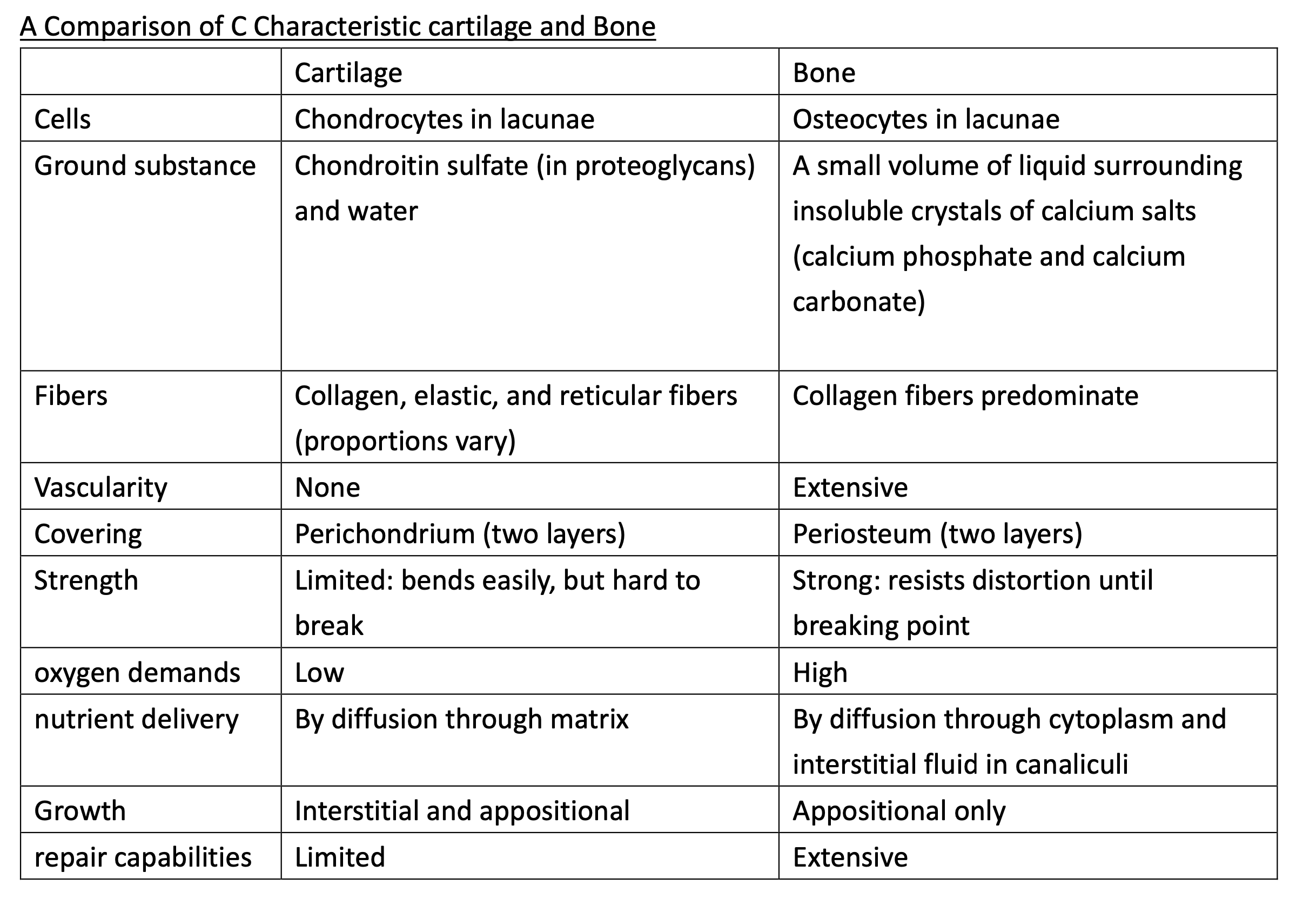
Supporting connective tissues
Cartilage
matrix: firm gel (proteoglycans)
cells: chondrocytes
lacunae: small chambers
avascular > all exchange of nutrients and waste products by diffusion through the matrix
perichondrium
have blood vessels > provide oxygen and nutrients to the underlying chondrocytes.
outer (fibrous region of dense irregular connective tissue) > support and protection and attaches the cartilage to other structures
inner (cellular layer) > the growth and maintenance of the cartilage
Cartilage growth
Interstitial growth: enlarges the cartilage from within.
Chondrocytes in the cartilage matrix divide, and the daughter cells produce additional matrix
Appositional growth: adds new layers of cartilage to the surface
Types of Cartilage
Hyaline cartilage (common)
FUNCTIONS: Tough, somewhat flexible > closely packed collagen fibers
LOCATIONS: connections between the ribs and the sternum
nasal cartilages > supporting cartilage, passageways of the respiratory tract
Elastic cartilage > yellowish color
Resilient and flexible (numerous elastic fibers)
FUNCTION: Provides support, but tolerates distortion without damage and returns to original shape
LOCATION: external flap (the auricle, or pinna) of the outer ear, a passageway to the middle ear cavity (the auditory tube), epiglottis, small cartilages (the cuneiform cartilages) in the larynx, or voice box.
articular cartilages, cover bone surfaces within joints > elbow and knee
Fibrocartilage
durable and tough (little substance, matrix)
FUNCTION: Resists compression; prevents bone-to-bone contact; limits movement
LOCATION: intervertebral discs
Bone
Fasciae (body wall)
connective tissue layers and wrappings that support and surround organs
Superficial Fascia (hypodermis, subcutaneous tissue)
CONTAINS: Areolar tissue and adipose tissue
LOCATION: Between skin and underlying organs
FUNCTION: provides insulation and padding, and lets the skin and underlying structures move independently
Deep Fascia
each layer all the fibers run in the same direction
CONTAINS: dense irregular connective tissue
LOCATION: Bound to capsules, tendons, and ligaments
FUNCTION: Forms a strong, fibrous internal framework
helps the tissue resist forces coming from many directions
Subserous Fascia
CONTAIN: Areolar tissue
LOCATION: Between serous membranes and deep fascia
Tissue membranes
Mucous Membranes
passageways and chambers > communicate with the exterior
FUNCTION: moist to reduce friction, facilitate absorption or secretion.
contain simple epithelia > perform absorptive or secretory functions
LOCATION: digestive, respiratory, reproductive, urinary tracts
Serous membranes
CONTAIN: Very thin, but firm attached to body wall and organs / sealed, internal cavities of the trunk > not open to the exterior
FUNCTION: serous fluid to minimize friction between the surfaces >keep moist and slippery
LOCATION: pleura, pericardium, peritoneum,
Cutaneous Membrane
CONTAIN: squamous epithelium, dense irregular connective tissue
FUNCTION: thick, waterproof, usually dry
LOCATION: skin
Synovial Membranes
CONTAIN: areolar tissue (matrix of interwoven collagen fibers, proteoglycans, glycoproteins)
FUNCTION: lubricated to keep friction from damaging the opposing surfaces
LOCATION: Adjacent bones (joints, articulations)
muscle tissue
Skeletal muscle tissue (striated voluntary muscle)
long, cylindrical, striated, multinucleate
partially repair after injury
CONTAIN: muscle fibers (very large muscle cells—up to 0.3 m), several hundred nuclei,
myosatellite cells (satellite cells)>produce new muscle fibers
FUNCTION: Moves or stabilizes the position of the skeleton; guards entrances and exits to the digestive, respiratory, and urinary tracts; generates heat; protects internal organs
LOCATION: Combined with connective tissues and neural tissue in skeletal muscles > adjacent skeletal muscle fibers - tied together by collagen and elastic fibers - attached tendon or aponeurosis.
Cardiac Muscle Tissue (striated involuntary muscle)
short, branched, striated, usually with a single nucleus
cells are interconnected by intercalated discs.
repair incomplete after damage
CONTION: Nuclei, Cardiac muscle cells (cardiocyte), Intercalated Discs, Striations
LOCATION: Heart
FUNCTION: Circulates, blood; maintains blood pressure
membranes are locked together by desmosomes, proteogly cans, and gap junctions.
Smooth Muscle tissue (nonstriated involuntary muscle)
short, spindle-shaped, nonstriated, with a single, central nucleus
regenerate after an injury
Contain: small, spindle-shaped, with tapering ends and a single, oval nucleus
LOCATION: Found in hollow organs > the walls of blood vessels and in digestive, respiratory, urinary, and reproductive organs
FUNCTION: Moves food, urine, and reproductive tract secretions; controls diameter of respiratory passageways; regulates diameter of blood vessels
Neural tissue
propagation (movement) of electrical impulses from one region of the body to another
control centers: brain and spinal cord
Neurons
Cell body: contains nucleus and major organelles
Dendrites: contacted by other neurons, receive information from other neurons
Axon: send information to other cells
Neuroglia (glial cells)
FUNCTION: support and repair neural tissue and supply nutrients to neurons
Reference
Martini, F. H., Nath, J. L., & Bartholomew, E. F. (2018). Fundamentals of anatomy & physiology (11th ed.). Pearson.
Contact Me
Instagram: https://www.instagram.com/weibuh_rn/
Threads: https://www.threads.net/@weibuh_rn
🌼 This note is my personal class note, no profit is made from sharing it
🌼 Whether it‘s exam applications or any questions during preparation, feel free to DM me on IG
🌼If there are errors in the content or copyright issues to contact via email: WeiBuh.RN@gmail.com