


✨ Nursing Note A&P – Osseous tissue and Bone Structure✨

看到一堆英文別怕別怕!!
因為許多人有考RN的需求,所以我手頭有筆記,就更新上來了
下篇預告:第一篇產科,胎心音!!!
Skeletal system
five primary functions
Support: provide a framework for the attachment of soft tissues and organs
Storage of Minerals and Lipids: Calcium is the most abundant mineral
skeleton store energy(lipids) in yellow bone marrow
Blood Cell Production: red bone marrow > RBC, WBC
Protection: surround many soft tissues and organs
Leverage: movements > precise motion, changes in the position
Bone
Bone Shapes
Sutural bones / Wormian bones: small, flat, irregularly shaped bones
Irregular bones: complex shapes. e.g., vertebrae, Skull (Zygomatic, sphenoid, ethmoid, maxilla, palatine, nasal hyoid, mandible, temporal), Pelvic bone, Sarum, Coccyx
Short bones: boxlike. e.g., Carpal(wrist), tarsal(ankle)
Flat bones: thin, flattened shape. e.g., Skull (Sphenoid, ethmoid, frontal, parietal, temporal, occipital), Scapula, Sternum, Ribs
Long bones: Long, slender. e.g., clavicle, Limbs (humers, ulna, radius, femur, tibia, fibula)
consist of a shaft (diaphysis) with two ends (epiphyses)
Sesamoid bones: small, flat, like sesame seed. (Figure 6–1f). e.g., patellae (kneecaps)
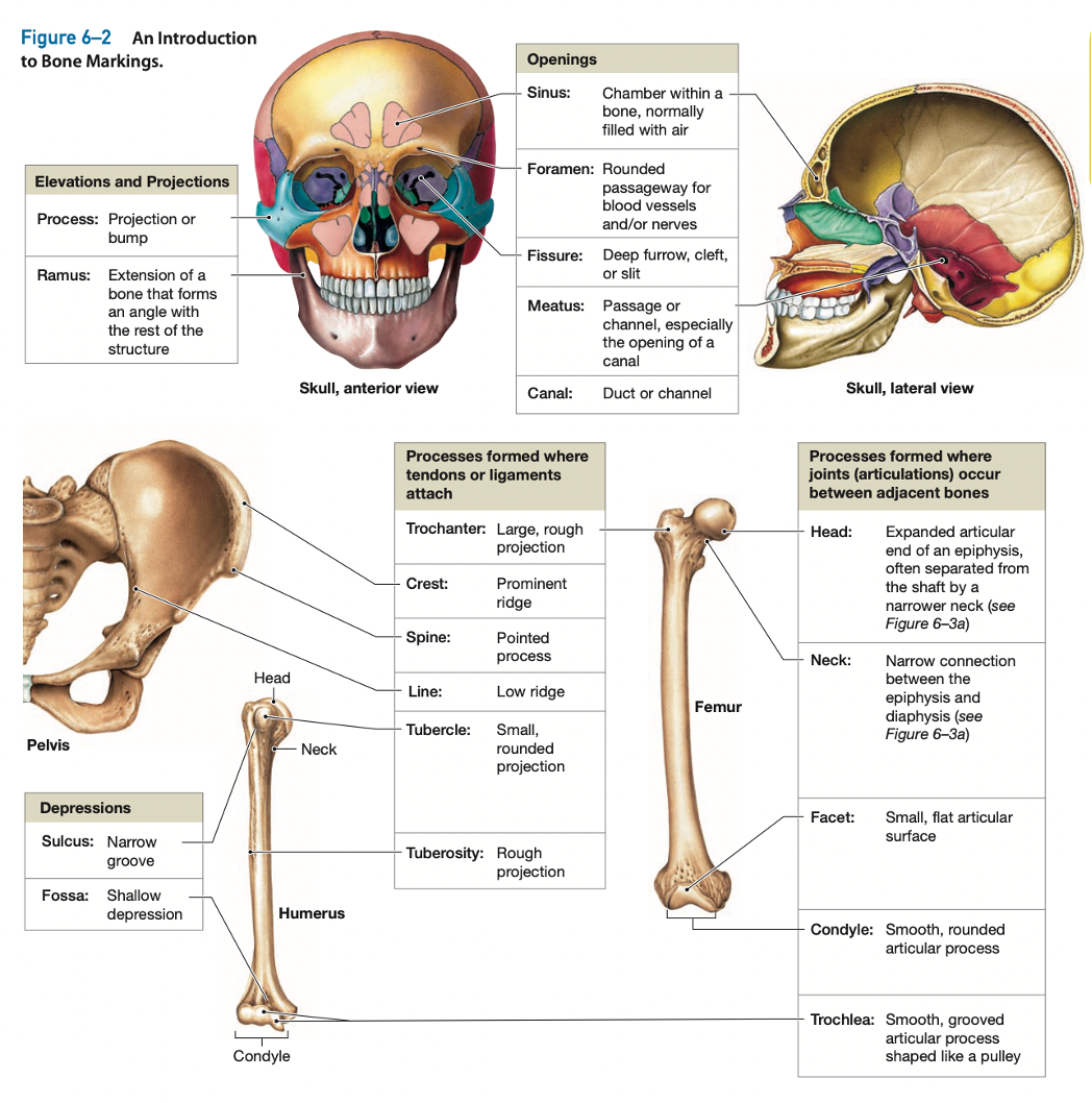
Bone Markings
Bone Structure

Bone Cells
Osteocytes(most): mature bone cells, cannot divide
Each osteocyte occupies a lacuna
FUNCTION:
maintain the protein and mineral content of the surrounding matrix
take part in the repair of damaged bone
Osteoblasts: produce new bone matrix (extracellular matrix, ECM), ossification / osteogenesis make and release the proteins and other organic components of the matrix(osteoid)
Osteogenic cell / osteoprogenitor cell: mesenchymal cells / stem cell > into osteoblasts
repair of a fracture
found in the inner, cellular layer of the periosteum
Osteoclasts: add matrix
50 or more nuclei
from monocytes and macrophages > body’s defense mechanisms.
produce acids and protein-digesting enzymes > dissolve the matrix and release the stored minerals.
Osteolysis: erosion process, resorption
regulation of calcium and phosphate concentrations in body fluids
removing matrix
Bone Matrix
Calcium phosphate (2/3), collagen fibers (1/3)
solid and sturdy >> calcium salts around the protein fibers
contains bone cells / osteocytes in lacunae
Compact bone
Periosteum: superficial layer > fibrous layer (outer) and cellular layer (inner)
FUNCTION:
(1) isolates the bone from surrounding tissues
(2) provides a route for the blood vessels and nerves
(3) takes part in bone growth and repair
Collagen fibers layer (outer): adjacent joint capsules, attached tendons and ligaments.
Osteons / Haversian system: cycle, osteocytes (cell) are arranged in concentric layers around a vascular central canal / Haversian canal
FUNCTION: protect, support, and resist stress.
Central canals: contains a capillary, a venule (very small vein) > carry blood to and from the osteon 水平的管路,開口在表面
Perforating canals / Volkmann’s canals: supply blood to osteons deeper in the bone and to tissues of the medullary cavity. 垂直的管路,在骨頭的中間
Lamellae: Surrounding the central canal are rings of bone ECM
give compact bone a great deal of strength,
Concentric lamellae: encircle the osteon.
Circumferential lamellae: encircle outermost
Interstitial lamellae: located between osteons
Lacunae: between the lamellae are small cavities, spindle-shaped spaces, around blood vessels
CONTAIN: osteocytes (mature osteoblasts)
Canaliculi (collagen fibers): narrow passageways between the lacunae > exchange of nutrients, wastes, gases (blood vessels)
Endosteum: connective tissue membrane (inner layer), incomplete cellular layer
CONTAIN: osteoblasts
FUNCTION: bone growth, repair, and remodeling

Spongy bone
Trabeculae: matrix in spongy bone forms a meshwork of supporting bundles of fibers
inside of a bone deep to compact bone,
FUNCTION: stores marrow
red bone marrow: (V) blood vessels, produce blood cells,
yellow bone marrow: composed primarily of adipose tissue (energy reserve)
no capillaries or venules in matrix
Bones formation and growth
Bone growth until about age 25
Ossification / osteogenesis: bone formation
Endochondral
Intramembranous
Calcification: deposition of calcium salts— takes place during ossification
Endochondral Ossification
bones originate hyaline cartilages > gradually replaced by bone
e.g., Long bones
STEPS
(1) cartilage enlarges
Enlarging chondrocytes within calcifying matrix
Disintegrating chondrocytes of the cartilage model > form cavities
(2) creating Primary ossification center
Blood vessels grow around the edges of the cartilage
perichodrium cell > convert to osteoblasts.
form Epiphysis, Diaphysis(shaft)
(3) Blood vessels penetrate the cartilage and invade the central region
Fibroblasts > migrating with the blood vessels differentiate into osteoblasts,
> begin producing spongy bone at a primary ossification center.
Bone formation > spreads along the shaft toward both ends of the former cartilage model.
(4) creating a medullary cavity
Diaphysis > becomes thicker, increases in length and diameter.
form Metaphysis
(5) creating secondary ossification centers
Capillaries and osteoblasts migrate into the epiphysis
(6) epiphysis filled with spongy bone
metaphysis> separates the epiphysis from the diaphysis.
shaft side > osteoblasts continuously invade the cartilage and replace it with bone.
epiphyseal side > New cartilage is produced
(7) Bone growth slow, then stop
At puberty: rate of epiphyseal cartilage production slows, and the rate of osteoblast activity accelerates.
At Adult: epiphyseal cartilage gets narrower and narrower, until it ultimately disappears. epiphyseal closure: the completion of epiphyseal growth > form epiphyseal line(生長停止線)
interstitial growth: continue to grow by expansion of the cartilage matrix
appositional growth: production of more cartilage at the outer surface
intramembranous Ossification / dermal ossification
begins when osteoblasts differentiate within a mesenchymal or fibrous connective tissue
takes place in the deeper layers of the dermis.
e.g., dermal bones/flat bones: skull, mandible (lower jaw), clavicles
Bone Blood and Nerve
Nutrient Artery and Vein
Most bones have only one nutrient artery and one nutrient vein
Femur > more than one of each
Vessels: enter the bone through passageways (nutrient foramina) in the diaphysis
large vessels branches: form smaller perforating canals and extend along the length of the shaft into the osteons of the surrounding compact bone
Metaphyseal Vessels
supply blood to the inner (diaphyseal) surface of each epiphyseal cartilage (being replaced by bone)
Periosteal Vessels
provide blood to the superficial osteons of the shaft
Periosteum
Lymphatics: collect lymph from branches that enter the bone and reach individual osteons by the perforating canals.
Sensory nerves: penetrate the compact bone with the nutrient artery to innervate the endosteum, medullary cavity, and epiphyses
Bone remodeling
throughout life
FUNCTION: bone maintenance > recycling and renewing bone matrix
INVOLVES: osteocytes, osteoblasts, osteoclasts
If removal is faster than replacement > bones weaken
If deposition predominates > bones strengthen
Exercise, hormones, nutrition
exercise effects on Bone
exercise > bone is stressed, mineral crystals generate minute electrical fields > Osteoblasts are attracted to these electrical fields >> begin to produce bone
inactivity > Degenerative changes in the skeleton
nutritional and hormonal effects on Bone
Minerals
(important)calcium and phosphate salts
magnesium, fluoride, iron, manganese
Calcitriol and cholecalciferol (vitamin D3)
expose sunlight > steroid cholecalciferol (vitamin D3) > synthesize calcitriol in the kidneys
absorbed from the diet
Vitamin C
absorbed from the diet
required for enzymatic reactions in collagen synthesis, stimulates osteoblast differentiation.
vitamin C deficiency: loss of bone mass and strength.
Vitamins A, K, B12
effects on bone structure
Vitamin A: stimulates osteoblast activity
Vitamins K and B12: synthesis of proteins in normal bone
Growth hormone
produced by the pituitary gland
stimulates protein synthesis and the rates of cell division and cell growth
Thyroxine
from the thyroid gland, stimulate bone growth
stimulates cell metabolism and increases the rate of osteoblast activity.
Sex hormones
At puberty
estrogens(females) and androgens (males)
stimulate osteoblasts to produce bone faster than the rate of epiphyseal cartilage expands
Calcium in bone physiology
(1) the bones (storage)
(2) the digestive tract (absorption)
(3) the kidneys (excretion)
Low Calcium Ion Level (<8.5 mg/dl)
Parathyroid Gland Response: secrete parathyroid hormone (PTH)
> Bone Response: Osteoclasts release stored calcium ions from bone
> Intestinal Response: increases intestinal absorption of calcium
> Kidney Response: Kidneys absorb calcium ions (decrease Calcium loss in urine)
>> Increase Calcium levels in blood
High Calcium Ion Level (>11 mg/dl)
Parathyroid Gland Response: Parafollicular cells (C cells) secrete calcitonin
> Bone Response: Osteoclasts inhibited to lock calcium ions in bone matrix
> Intestinal Response: decreases intestinal absorption of calcium
> Kidney Response: Kidneys allow calcium loss (Increase Calcium loss in urine)
>> decrease Calcium levels in blood
Reference
Martini, F. H., Nath, J. L., & Bartholomew, E. F. (2018). Fundamentals of anatomy & physiology (11th ed.). Pearson.
Contact Me
Instagram: https://www.instagram.com/weibuh_rn/
Threads: https://www.threads.net/@weibuh_rn
🌼 This note is my personal class note, no profit is made from sharing it
🌼 Whether it‘s exam applications or any questions during preparation, feel free to DM me on IG
🌼If there are errors in the content or copyright issues to contact via email: WeiBuh.RN@gmail.com